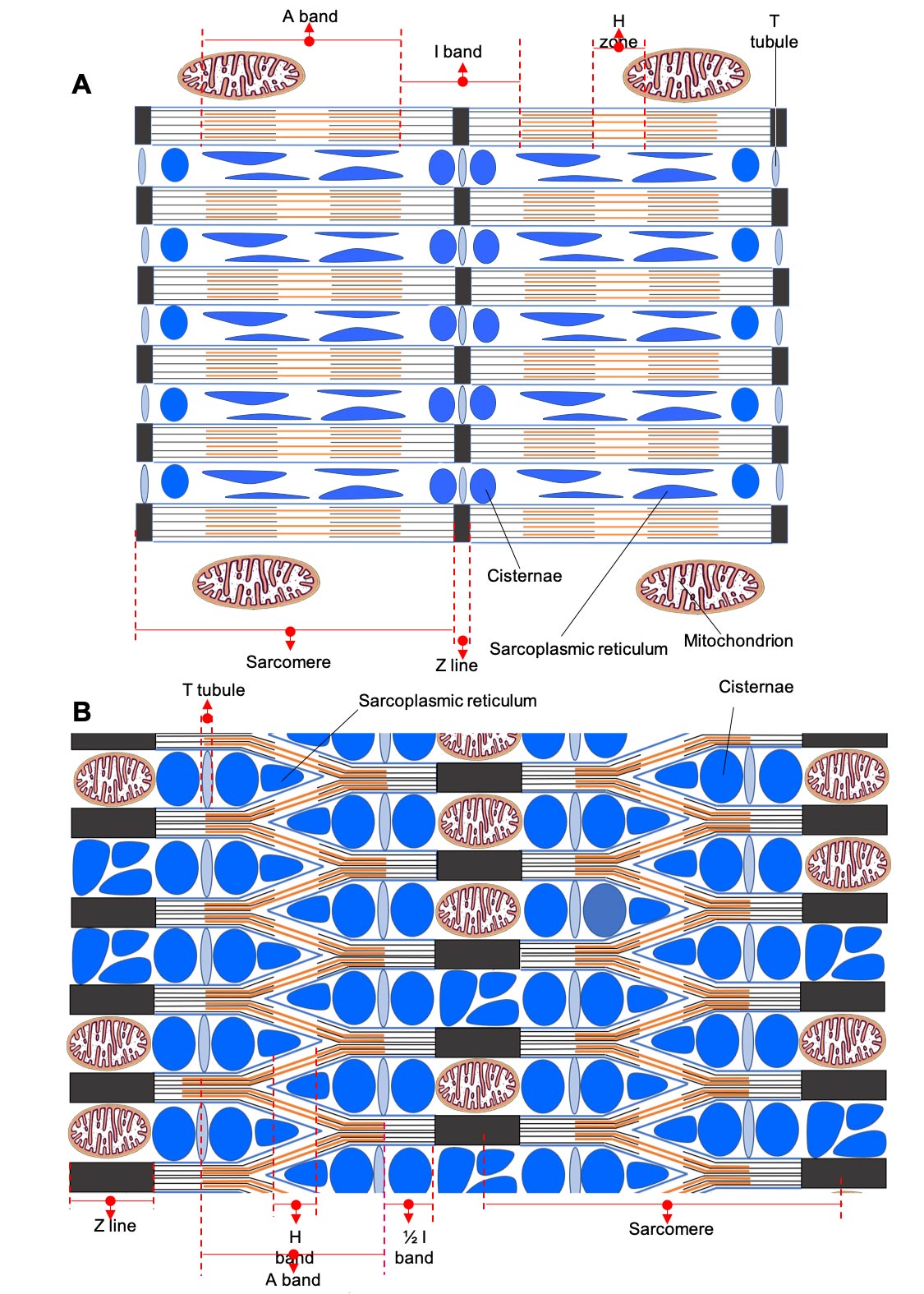
Langsdoorsnede die de netachtige formatie in de stemspier in V Parovedion Vassali. Credits: Marc Thierry/Universiteit van Luik
Onderzoekers hebben zojuist een onverwachte ontdekking gedaan van een nieuwe organisatie van spiervezels in Parovedion Vassali, een vis die in de Middellandse Zee leeft en, zoals veel vissen, gespecialiseerde spieren gebruikt om geluiden te maken. Dit is een belangrijke ontdekking die ons begrip van spiercontractie zou kunnen veranderen.
Wetenschappers van de Universiteit van Luik, Eric Parmentier en Marc Thierry, hebben een unieke opstelling van spiervezels ontdekt in de mediterrane vis, Parophidion vassali, die ons begrip van spiercontractie radicaal kan veranderen. Deze unieke mesh-achtige configuratie van spiervezels in een spiervezel kan snelle samentrekkingen mogelijk maken met behoud van kracht. Er is meer onderzoek nodig om de nieuwe spiervezelstructuur en de functionele implicaties ervan volledig te begrijpen.
De beschrijving van skeletspieren dateert uit observaties van de Nederlandse bioloog Antonie van Leeuwenhoek, een inleiding tot celbiologie en microbiologie die in een artikel gepubliceerd in 1712 in de Philosophical Transactions of the Royal Society bedankte. Met behulp van een draagbare microscoop met één lens, de eerste beschrijving van spiervezels van een walvis. In 1840 gaf anatoom William Bowman een nauwkeurigere beschrijving van de spier en merkte op: “de aanwezigheid en plaatsing van afwisselend lichte en donkere lijnen […] Die zich kenmerkt door precisie en verfijnde afwerking.” (Zie over spiervezels onderaan dit artikel.)
Latere studies hebben geleid tot duidelijkere beschrijvingen, waaronder identificatie van de verschillende moleculen waaruit spieren bestaan en verklaringen van hoe ze functioneren, in het bijzonder het model van spiercontractie voorgesteld door biofysicus Andrew Huxley in 1957. In de afgelopen 300 jaar zijn veel onderzoeken uitgebreid de weergave van zijn regulering Spiervezels in verschillende klassen. Dit toonde aan dat de algemene organisatie van dwarsgestreepte spiervezels volledig behouden was in alle tot nu toe bestudeerde groepen gewervelde dieren.

Figuur 1. Vergelijking van ‘klassieke’ skeletspierorganisatie met parallelle myofibrillen en stemspier met reticulaire myofibrillen in Parovedion Vassali. Credits: Parmentier/M. Terry/Universiteit van Luik
De verhouding van elk van deze cellulaire componenten kan echter variëren van vezel tot vezel, waardoor deze vezels speciale samentrekkende eigenschappen hebben. Vezels die rijk zijn aan myofibrillen met een slecht ontwikkeld sarcoplasmatisch reticulum worden bijvoorbeeld aangetroffen in spieren die kracht ontwikkelen tijdens samentrekking. Integendeel, er zijn verminderde vezels in myofibrillen met een overvloed aan sarcoplasmatisch reticulum en veel mitochondriën in spieren die een verhoogde contractiesnelheid ontwikkelen. De snelste spieren zijn te vinden in de stemspieren van vissen, waar ze nadrukkelijk aanwezig zijn[{” attribute=””>species produce sounds using muscles that contract at a frequency of between 100 and 300 Hz, i.e. 100 to 300 contraction/relaxation cycles per second.
A recent study carried out in collaboration between the Functional and Evolutionary Morphology Laboratory, and the Cell and Tissue Biology Laboratory has revealed a new arrangement of myofibrils within the fibers of a sonic muscle in the fish Parophidion vassali. Rather than being arranged in parallel, the myofibrils form an enormous network within the muscle fiber, explains Prof Eric Parmentier, director of the Functional and Evolutionary Morphology Laboratory at the University of Liège.
Each myofibril subdivides into two branches at each sarcomere, one connecting to the myofibril above and the other connecting to the myofibril below (Figure 1B). This new muscle fiber design could result in a muscle that contracts rapidly while retaining strength. The paucity of myofibrils and the high volume occupied by the sarcoplasmic reticulum are in favor of fibers that contract rapidly.
“The network structure of the myofibrils would allow more myosin heads to form cross-bridges with the actin myofilaments, which would increase strength in this fast muscle,” explains Prof Marc Thiry, Director of the Cell and Tissue Biology Laboratory. “In addition, numerous mitochondria unusually arranged within the Z striae (very long in these fibers: 700 nm compared with 70 to 150 nm in a conventional muscle) appear to provide the energy needed to produce long-lasting sounds.”
This new type of organization of striated muscle fibers, never before described in the scientific literature, and which would therefore make it possible to combine muscular strength and speed, requires further studies to understand how it works and to determine whether there are adaptations at the level of the different molecules involved in these muscles.

Longitudinal section in a classic skeletal muscle. Credit: Marc Thiry/University of Liège
About Muscle Fibers
Striated skeletal muscle fibers or cells represent the elementary units of voluntary muscles (muscles that enable movements such as locomotion or posture maintenance) in animals. “Each fiber is characterized by numerous contractile elements, actin, and myosin myofilaments, organized in bundles parallel to the muscle fiber’s long axis, called myofibrils.
In the longitudinal section under the optical microscope, these fibers appear as a succession of light and dark bands located at the same level for each myofibril, giving the appearance of transverse striation to the muscle fiber. A darker line divides the light band in the middle, known as the Z striation. The portion of the myofibril between two Z striae is called a sarcomere and represents the contractile unit of the myofibril. Each myofibril is therefore made up of many sarcomeres placed end to end.
The myofibrils occupy a large cell volume and are surrounded by cisternae of the smooth endoplasmic reticulum (or sarcoplasmic reticulum), which stores the calcium essential for muscle contraction. In addition, mitochondria are located close to the myofibrils; they are the main source of ATP providing energy for muscle contraction.

“Reizende ninja. Onruststoker. Spekonderzoeker. Expert in extreme alcohol. Verdediger van zombies.”


More Stories
Griepseizoen vliegt voorbij met verkoudheid – DiscoverMooseJaw.com
De eeuwenoude overblijfselen die in een put zijn gevonden, kunnen van een man uit de Noorse sagen zijn
Een ruimtetoerist raakt van streek als het zicht begint te verslechteren